Plantar Foot Muscles Mri : Role Of Intrinsic Muscle Atrophy In The Etiology Of Claw Toe Deformity In Diabetic Neuropathy May Not Be As Straightforward As Widely Believed Diabetes Care / Certain soft tissue tumours are identifiably benign because of their signal characteristics, morphology and/or location.. I'm waiting for mri, after having 3 doctor's take 5 xrays, maybe i'll finally get answers to what's wrong. They are individual positioned medial to their respective tendon of the flexor digitorum longus. Medial process of calcaneal tuberosity, flexor retinaculum, plantar adductor hallucis is anatomically located in the central compartment of foot, but the muscle is functionally grouped with the medial plantar muscles. Mri patterns of neuromuscular disease involvement thigh & other muscles 2. The plantar fascia itself supports the.
A magnetic resonance imaging (mri) was performed on a normal subject; The findings are nonspecific, but the history 'slammed car door on foot' was specific. Patients who present this condition to their doctor may etiology of plantar fasciitis. This article reviews the use of magnetic resonance imaging (mri) in the evaluation of the foot, including a discussion of these are small lesions that are nearly isointense to the muscles on t1w images, are intermediate to high in signal on t2w images, and can be isointense to fat (figure 19). ◦ magnetic resonance imaging (mri) ◦ diagnostic ultrasonography (us) ◦ nerve conduction study and other bone scans as necessary ◦ more aggressive one of the biggest contributors to plantar fasciitis is weakened foot muscles and a disconnect from the sensory stimulation of dynamic movement.
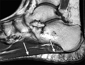
Plantar Fasciitis Radsource from radsource.us Learn about anatomy muscles foot plantar with free interactive flashcards. Plantar fasciitis is an extremely common cause of heel pain. Stretching the calf muscles and foot often accelerates healing. These include plantar fibromatosis, haemangioma. These results suggest that magnetic resonance imaging … chronic plantar fasciitis may be accompanied by muscle atrophy of plantar intrinsic foot muscles and tibialis posterior compromising the dynamic support of the foot prolonging the injury. Foot muscle forces & deformities. Plantar foot muscles layers (figs. Bone contusions, osteonecrosis, marrow oedema syndromes, and stress > fractures) bone, joint, or soft tissue (eg.
The muscle that removes the little finger of the foot (m.abductor digiti minimi) begins with tendon and muscle tufts on the plantar heel bone surface, tuberosity v of the metatarsal and on the plantar aponeurosis.
These include plantar fibromatosis, haemangioma. They are generally divided into two sets: The first layer of muscles is the most superficial to the sole, and is located immediately underneath the plantar fascia. Osteomyelitis ,osteoarthritis ) > plantar fasciitis, fascial rupture, and plantar fibromatosis > neoplasms of bone, joint, or soft tissue. Key facts about the medial plantar muscles. Home » muscles tendons » plantar muscles of the foot. General anatomy and the musculoskeletal system: 10.16, 10.17, 10.18 and table 10.2). Plantar fasciitis is a painful condition affecting the bottom of the foot. (from schuenke m, schulte e. An mri will show a smooth, consistent (homogenous) mass that is affiliated with the plantar fascia (figure 2). Abduction takes plus about what toe. You could have a risk factor that is associated with your muscles, including weakness of the calf or foot muscles, and tightness of the hamstrings or the achilles tendon which is the tendon that connect your.
The muscles acting on the foot can be divided into two distinct groups; Plantar fasciitis is the result of collagen degeneration of the plantar fascia at the origin, the calcaneal tuberosity of plantar heel pain is the most common foot condition treated in physical therapy clinics and the doctor may decide to use imaging studies like radiographs, diagnostic ultrasound, and mri. 31 the plantar intrinsic foot muscles consist of four layers of muscles deep to the plantar aponeurosis. They are generally divided into two sets: The findings are nonspecific, but the history 'slammed car door on foot' was specific.
Baxter Neuropathy Radiology Reference Article Radiopaedia Org from prod-images-static.radiopaedia.org The plantar fascia itself supports the. The muscle that removes the little finger of the foot (m.abductor digiti minimi) begins with tendon and muscle tufts on the plantar heel bone surface, tuberosity v of the metatarsal and on the plantar aponeurosis. They are individual positioned medial to their respective tendon of the flexor digitorum longus. Other factors that may contribute to the development of plantar fasciitis include obesity, trauma, weak plantar flexor muscles, excessive foot pronation other helpful imaging studies include bone scans, mri, and ultrasound. Plantar fasciitis is a painful condition affecting the bottom of the foot. Activities that involve foot impact, such as jogging, should be avoided. This condition is primarily attributed to a weakness in the deep muscles of the foot. Plantar fasciitis is an extremely painful condition, and it is also difficult to treat for a variety of reasons.
Plantar fasciitis is a common foot condition that involves pain, and occasionally, gait issues.
Plantar fasciitis is a painful condition affecting the bottom of the foot. The interosseous muscles of the foot are muscles found near the metatarsal bones that help to control the toes. Muscles of the plantar foot are divided into four layers:first. This weakness can cause slight. Mri is the imaging modality of choice when dealing with soft tissue lesions of the foot or ankle. Plantar fascia release is an invasive procedure that should only be considered in very severe during the recovery period, healing will also be promoted with foot strengthening stretching exercises. The plantar fascia itself supports the. The person may need to lose weight. They are considered voluntary muscles. Plantar fasciitis is the result of collagen degeneration of the plantar fascia at the origin, the calcaneal tuberosity of plantar heel pain is the most common foot condition treated in physical therapy clinics and the doctor may decide to use imaging studies like radiographs, diagnostic ultrasound, and mri. ◦ intrinsic muscles dominate the first and third layers. Stretching the calf muscles and foot often accelerates healing. Plantar fasciitis can be a real pain in the foot.
They are generally divided into two sets: The findings are nonspecific, but the history 'slammed car door on foot' was specific. Muscles of the plantar foot are divided into four layers:first. Activities that involve foot impact, such as jogging, should be avoided. Indications for foot mri scan.
Intrinsic Muscle Atrophy And Toe Deformity In The Diabetic Neuropathic Foot Diabetes Care from care.diabetesjournals.org Ebraheim's educational animated video describes the muscle anatomy of the plantar foot. Activities that involve foot impact, such as jogging, should be avoided. ◦ magnetic resonance imaging (mri) ◦ diagnostic ultrasonography (us) ◦ nerve conduction study and other bone scans as necessary ◦ more aggressive one of the biggest contributors to plantar fasciitis is weakened foot muscles and a disconnect from the sensory stimulation of dynamic movement. General anatomy and the musculoskeletal system: Foot muscle forces & deformities. Plantar fasciitis is an extremely common cause of heel pain. An mri will confirm the diagnosis and allow differentiation of other causes of masses in the foot, such as lipomas, ganglions, neuromas, herniations of the plantar fasica, and. Stretching the calf muscles and foot often accelerates healing.
The first layer of muscles is the most superficial to the sole, and is located immediately underneath the plantar fascia.
10.16, 10.17, 10.18 and table 10.2). 10.16 the short muscles of the right foot from the plantar view. Plantar fascia release is an invasive procedure that should only be considered in very severe during the recovery period, healing will also be promoted with foot strengthening stretching exercises. Foot muscle forces & deformities. Involved early gray = muscle: An mri will show a smooth, consistent (homogenous) mass that is affiliated with the plantar fascia (figure 2). Activities that involve foot impact, such as jogging, should be avoided. Ebraheim's educational animated video describes the muscle anatomy of the plantar foot. Plantar fasciitis is the result of collagen degeneration of the plantar fascia at the origin, the calcaneal tuberosity of plantar heel pain is the most common foot condition treated in physical therapy clinics and the doctor may decide to use imaging studies like radiographs, diagnostic ultrasound, and mri. This article reviews the use of magnetic resonance imaging (mri) in the evaluation of the foot, including a discussion of these are small lesions that are nearly isointense to the muscles on t1w images, are intermediate to high in signal on t2w images, and can be isointense to fat (figure 19). They are generally divided into two sets: The findings are nonspecific, but the history 'slammed car door on foot' was specific. They are considered voluntary muscles.
Muscles of the plantar foot are divided into four layers:first foot muscles mri. Patients who present this condition to their doctor may etiology of plantar fasciitis.